


 With advancements in 3D printing, surgeons can now use the technology to produce replicas of baby’s hearts in order to better understand what needs to be fixed.
Rare congenital disorders in newborns can create situations where the heart is malformed, however the tiny size of the heart and the child make surgeries difficult and often dangerous. Having a 3D roadmap of the heart before going in for the surgery can act as an indispensable tool for surgeons to keep the surgery as uncomplicated and quick as possible.
Researchers presented findings from using replica hearts November 19, 2014 at the American Heart Association meeting in Chicago, reports Live Science. Though very few of the replicas have been used so far each has shown great promise in providing superior tools for heart surgeons.
Study co-author Dr. Matthew Bramlet said to Live Science, “From the first two cases straight out of the gate, we’ve had this dramatic impact.” Bramlet is a pediatric cardiologist at the University of Illinois College of Medicine and the Children’s Hospital of Illinois, both in Peoria.
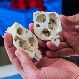
Bramlet used detailed MRI scans do design anatomically accurate models of hearts and they were printed at the Jump Trading Simulation and Education Center, also in Peoria.
In the first listed case doctors believed the child had a single hole in one wall of the heart; however, the printed heart, based on MRI, revealed further holes that needed to be closed. With a roadmap of all the holes and their locations, doctors were far better prepared for the surgery.
In the second case, the baby had such a hole and several defects with arteries leading to the heart. The process to fix these sorts of defects is complex and arduous, and as result very stressful for the patient. However, after holding the replica heart the surgeon was able to determine a safer, less-complex surgery.
According to Live Science since the first repair using a model heart, the team has gone on to create eight or nine heart replicas. Every single one improved the surgeons understanding of the future surgery.
It is cautioned that so far too few heart replicas have been produced to know if this is a viable tool. Part in fact because complicated heart defects are rare and 3D printing is still a growing technology. As a result, it’s hard to find cases that it is applicable to—but so far it seems that when a replica heart is called for it provides clues that could otherwise be lost with standard imaging and visualization technology.
Live Science has further photos and commentary on the replica hearts and the replication process.
Support our mission to keep content open and free by engaging with theCUBE community. Join theCUBE’s Alumni Trust Network, where technology leaders connect, share intelligence and create opportunities.
Founded by tech visionaries John Furrier and Dave Vellante, SiliconANGLE Media has built a dynamic ecosystem of industry-leading digital media brands that reach 15+ million elite tech professionals. Our new proprietary theCUBE AI Video Cloud is breaking ground in audience interaction, leveraging theCUBEai.com neural network to help technology companies make data-driven decisions and stay at the forefront of industry conversations.